Home

פסטה מורדרין קו אורך lamina cribrosa הרם מערך ריקבון

High resolution in vivo imaging of the lamina cribrosa - ScienceDirect

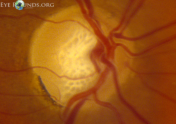
Atlas Entry - Lamina cribrosa in primary open angle glaucoma

Cribriform plate (Lamina cribrosa); Image: Yousun Koh | Skull anatomy,  Bones, Sphenoid bone
Cribriform plate (Lamina cribrosa); Image: Yousun Koh | Skull anatomy, Bones, Sphenoid bone

Ethmoid bone: Anatomy, borders and development | Kenhub

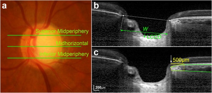
Ocular and Clinical Characteristics Associated with the Extent of Posterior Lamina  Cribrosa Curve in Normal Tension Glaucoma | Scientific Reports
Ocular and Clinical Characteristics Associated with the Extent of Posterior Lamina Cribrosa Curve in Normal Tension Glaucoma | Scientific Reports

Using OCT to Measure Anterior Lamina Cribrosa Depth - Glaucoma Today

Lamina cribrosa pore movement during acute intraocular pressure rise |  British Journal of Ophthalmology
Lamina cribrosa pore movement during acute intraocular pressure rise | British Journal of Ophthalmology

Location of lamina cribrosa in the optic nerve. | Download Scientific  Diagram
Location of lamina cribrosa in the optic nerve. | Download Scientific Diagram

Determinants of lamina cribrosa depth in healthy Asian eyes: the Singapore  Epidemiology Eye Study | British Journal of Ophthalmology
Determinants of lamina cribrosa depth in healthy Asian eyes: the Singapore Epidemiology Eye Study | British Journal of Ophthalmology

Ethmoid bone
Ethmoid bone

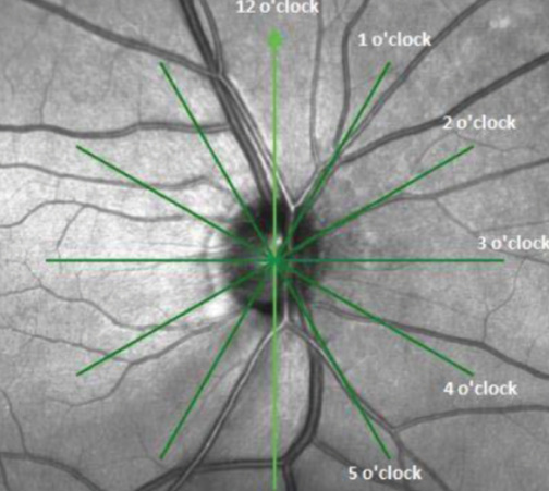
Focal Lamina Cribrosa Defect in Myopic Eyes With Nonprogressive  Glaucomatous Visual Field Defect - American Journal of Ophthalmology
Focal Lamina Cribrosa Defect in Myopic Eyes With Nonprogressive Glaucomatous Visual Field Defect - American Journal of Ophthalmology

A biomathematical model for pressure-dependent lamina cribrosa behavior -  ScienceDirect
A biomathematical model for pressure-dependent lamina cribrosa behavior - ScienceDirect

File:Cranium - os ethmoidale, lamina cribrosa (superior view, detail).jpg -  Wikimedia Commons
File:Cranium - os ethmoidale, lamina cribrosa (superior view, detail).jpg - Wikimedia Commons

Using OCT to Measure Anterior Lamina Cribrosa Depth - Glaucoma Today

Os Ethmoidale Flashcards | Quizlet

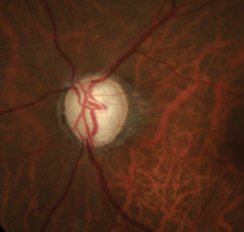
PLOS ONE: Lamina Cribrosa Defects and Optic Disc Morphology in Primary Open  Angle Glaucoma with High Myopia
PLOS ONE: Lamina Cribrosa Defects and Optic Disc Morphology in Primary Open Angle Glaucoma with High Myopia

Reza Zadeh on Twitter: "Nerve fiber layer thickness is used as a metric to  track Glaucoma. The Lamina Cribrosa is not assigned with any metrics,  because clinicians don't have strongly correlated metrics
Reza Zadeh on Twitter: "Nerve fiber layer thickness is used as a metric to track Glaucoma. The Lamina Cribrosa is not assigned with any metrics, because clinicians don't have strongly correlated metrics
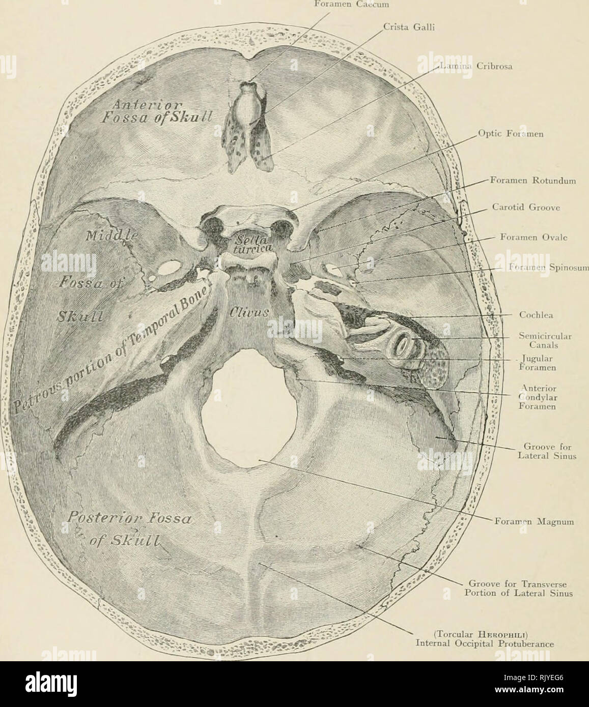
Cribrosa High Resolution Stock Photography and Images - Alamy

Atlas of applied (topographical) human anatomy for students and  practitioners. Anatomy. Lamina Cribrosa. Groove for Lateral Sinus Foramen  Magnum Groove for Transverse Portion of Lateral Sinus (Torcular Hbhophili)  Internal Occipital Protuberance
Atlas of applied (topographical) human anatomy for students and practitioners. Anatomy. Lamina Cribrosa. Groove for Lateral Sinus Foramen Magnum Groove for Transverse Portion of Lateral Sinus (Torcular Hbhophili) Internal Occipital Protuberance

PLOS ONE: Defects of the Lamina Cribrosa in High Myopia and Glaucoma

Unmyelinated portion of the optic nerve extending into the lamina... |  Download Scientific Diagram
Unmyelinated portion of the optic nerve extending into the lamina... | Download Scientific Diagram

International Glaucoma Review

The role of lamina cribrosa tissue stiffness and fibrosis as fundamental  biomechanical drivers of pathological glaucoma cupping | American Journal  of Physiology-Cell Physiology
The role of lamina cribrosa tissue stiffness and fibrosis as fundamental biomechanical drivers of pathological glaucoma cupping | American Journal of Physiology-Cell Physiology

Imaging of the lamina cribrosa and its role in glaucoma: a review - Tan -  2018 - Clinical & Experimental Ophthalmology - Wiley Online Library
Imaging of the lamina cribrosa and its role in glaucoma: a review - Tan - 2018 - Clinical & Experimental Ophthalmology - Wiley Online Library

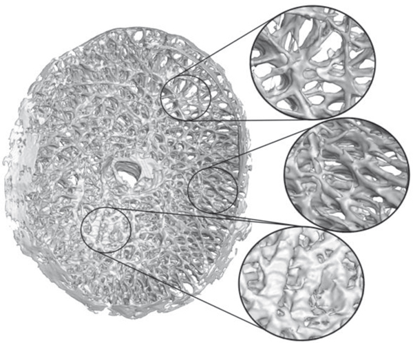
Figure 1 from Morphometry of the human lamina cribrosa surface. | Semantic  Scholar
Figure 1 from Morphometry of the human lamina cribrosa surface. | Semantic Scholar

Border Tissue Morphology Is Spatially Associated with Focal Lamina Cribrosa  Defect and Deep-Layer Microvasculature Dropout in Open-Angle Glaucoma -  American Journal of Ophthalmology
Border Tissue Morphology Is Spatially Associated with Focal Lamina Cribrosa Defect and Deep-Layer Microvasculature Dropout in Open-Angle Glaucoma - American Journal of Ophthalmology

HLS [ Eye, eye, lamina cribrosa] MED MAG